The Transparent Front Surface of the Eye Is Called the
Also the space between the cornea and the lens is filled with a gel-like fluid called the aqueous humor. It is the raised clear area directly in front of the iris.

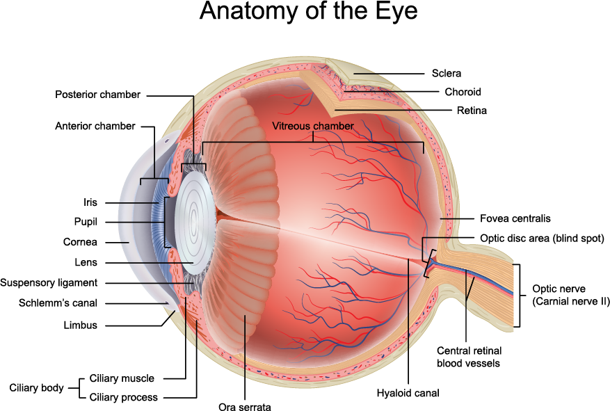
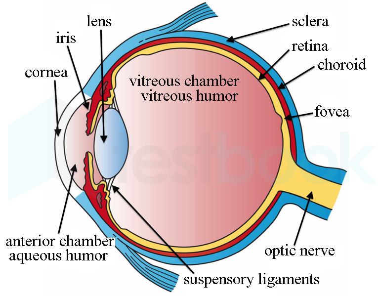
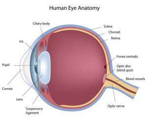
Human Eye Ball Anatomy Physiology Diagram
Like the glass on a watch the cornea is the clear protective coating on the front of the eye that allows light to pass through it without distortion.

. It is a powerful refracting surface providing 23 of the eyes focusing power. It is the eyes primary light-focusing structure. It covers the pupil iris and anterior chamber the fluid-filled inside of the eye.
A Transparent front part of eye ii Pupil b Layer on which impression of images is formed iii Iris c Point on retina where there are no nerve endings iv Retina d Sensitive for bright light v Blind spot e Is a small opening in the cornea vi Rods f Sensitive for dim light vii Cones g Controls the size of the pupil. The outer layer contains the sclera the white of the eye and the cornea the clear dome at the. It is avascular no vessels as its nourishment comes primarily from the air and from the tears.
Viewed from the front of the eye the cornea appears slightly wider than it is tall. Primary refractive surface of the eye. The cornea is a transparent avascular tissue that acts as a structural barrier and protects the eye against infections.
Light first enters the eye through a transparent structure on the surface of the eye called a pupil e. The clear front of your eye is called the cornea. Cornea contributes to two-third of the refractive power of the eye.
The angle made by a reflected ray with a perpendicular to the reflecting surface. The clear transparent tissue that is located on the very front anterior portion of the eye. The front surface of the eye is covered by the transparent.
This is because the sclera the white of the eye slightly overlaps the top and bottom of the anterior cornea. The front clear surface of the eye is called the cornea. The transparent front surface of the eye is called the a.
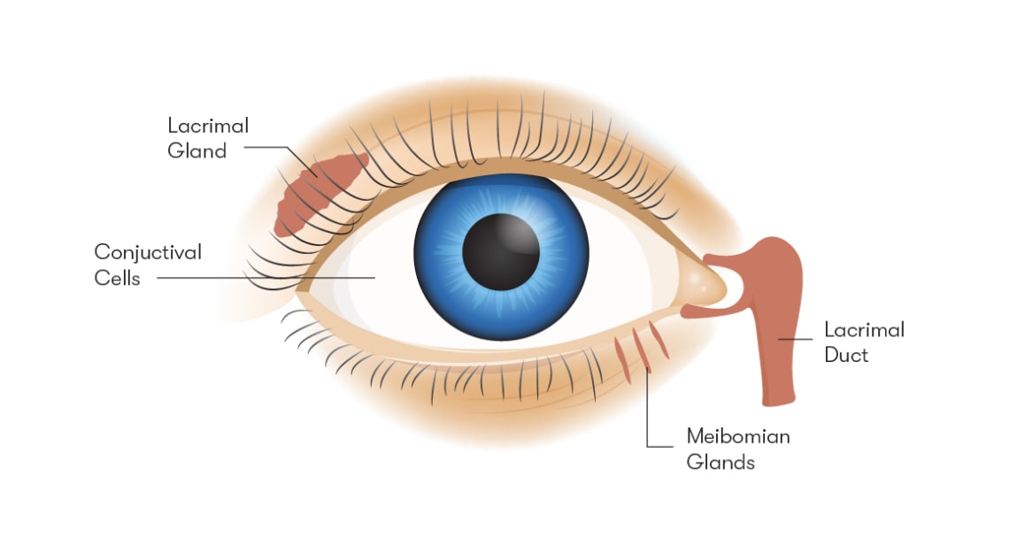
Tears lubricate the eye and are made up of three layers. The ___ protects the eye. The lens of the eye focuses the images transmitted through the cornea to the retina.
The outer transparent structure at the front of the eye that covers the iris pupil and anterior chamber. 1 Along with the tear film it provides proper anterior refractive surface for the eye. Like the crystal on a watch it gives us a clear window to look through.
The iris is partly responsible for regulating the amount of light permitted to enter the eye. It is the main refractive surface of the eye. The process by which waves can bend around corners or pass through openings.
The signals generated by the rods and cones are carried to your. The layers of the tear film keep the front of the eye lubricated. It is transparent in nature.
The colored ring of muscle that controls the size of the pupil is called the a. The clear dome-shaped surface that covers the front of the eye. Now up your study game with Learn mode.
One-sixth of the outer layer of the eye called the tunic fibrosa or fibrous tunic bulges forward as the cornea the transparent dome which serves as the outer window of the eyeThe cornea is the primary most powerful structure focusing light entering the eye along with the secondary focusing structure the crystalline lens. You just studied 23 terms. The cornea is the clear bulging surface in front of the eye.
Waves Light and Eye Review. The clear cellophane-like tissue that covers the sclera and the inside surface of eyelids. It is highly sensitive.
The coloured part of your eye is called the iris. Since it covers the pupil as well it is necessarily transparent so that we can see through it. It lies directly in front of the iris and pupil and it allows light to enter the eye.
Iris Colored tissue lying behind the cornea that gives color to the eye eg blue eyes brown eyes and controls the amount of light entering the eye by varying size of the black pupillary opening. The surface of the eye and the inner surface of the eyelids are covered with a clear membrane called the conjunctiva. Richly supplied with nerve fibers.
This transparent disc sits over the pupil and iris protecting them and letting in light. The cornea is the transparent part of the eye that covers the front portion of the eye. N 137.
The cornea also forms the first part of the process of focusing what you look at into an image on the back of your eye see below. Lens also called crystalline lens. The cornea is composed for the most part of.
The cornea is the clear front surface of the eye. Rods and cones are the light-senstive cells on the a. The colored part of the eye.
The transparent structure inside the eye that focuses light rays onto the retina. The palpebral conjunctiva lines the lids while the bulbar conjunctiva covers the sclera. It covers the colored iris.
Light enters the eye through the transparent front surface called the ___. Although the eyes pupil is indeed a hole in the front surface of the eye this hole is covered in the front by a strong transparent coating called the cornea and in the back by a fibrous transparent object called the lens. The b comea retina d iris.
Light enters the eye through a curved front surface known as cornea. It protects the covering of the eye and helps to focus light. Subsequently question is what is the outer layer of the eyeball called.
Deposits of yellowish extra cellular waste products that accumulate within and beneath the retinal pigmented epithelium RPE layer. The cornea is the other transparent covering. Normally transparent and uniformly thick.
The cornea is the transparent dome-shaped window covering the front of the eye. The transparent front segment of the eye that covers the iris pupil and anterior chamber and provides most of the eyes optical power.
Anatomy Of The Human Eye Pinpointeyes
What Is The Name Of A The Curved Transparent Front Tutorix

Pin By Siv K On My Notes Human Eye Eyes Lens

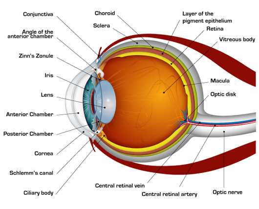
Eye Anatomy Bell Booth Sirkka Fabris Optometrists

4 Ways Cosmetic Dentistry Can Fix Crooked Teeth Cosmetic Dentistry Crooked Teeth Cosmetic Bonding
Eye Anatomy Retina Specialists Orlando Central Florida Retina
How The Human Eye Works Cornea Layers Role Light Rays

Christmas Tree Cataracts Are Found In Higher Prevalence In Patients With Myotonic Dystrophy Eye Facts Eye Vitamins Myotonic Dystrophy
Transparent Front Part Of Eye Is
What Is The Name For The Transparent Outer Covering Of The Front Of The Eye Quora

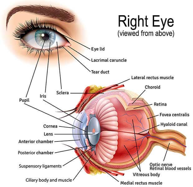
Eye Structures Front And Side Views

Eye Anatomy Bell Booth Sirkka Fabris Optometrists

Front Eye Level Perspective View Of The Black Ds9208 On A Glossy Surface And Grey White Gradient Background Design Sketch A Day Style Definition

Eye Anatomy Bell Booth Sirkka Fabris Optometrists
Solved The Transparent Front Part Of The Eye Is Called
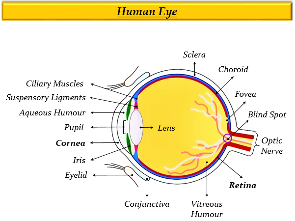
Parts Of The Eye
What Is The Name For The Transparent Outer Covering Of The Front Of The Eye Quora

First Surface Mirror Order High Precision Mirrors For Engineering Two Way Mirror Surface Mirror
The Eye Anatomy Vision Pro Optical Eye Care You Can Trust


Comments
Post a Comment